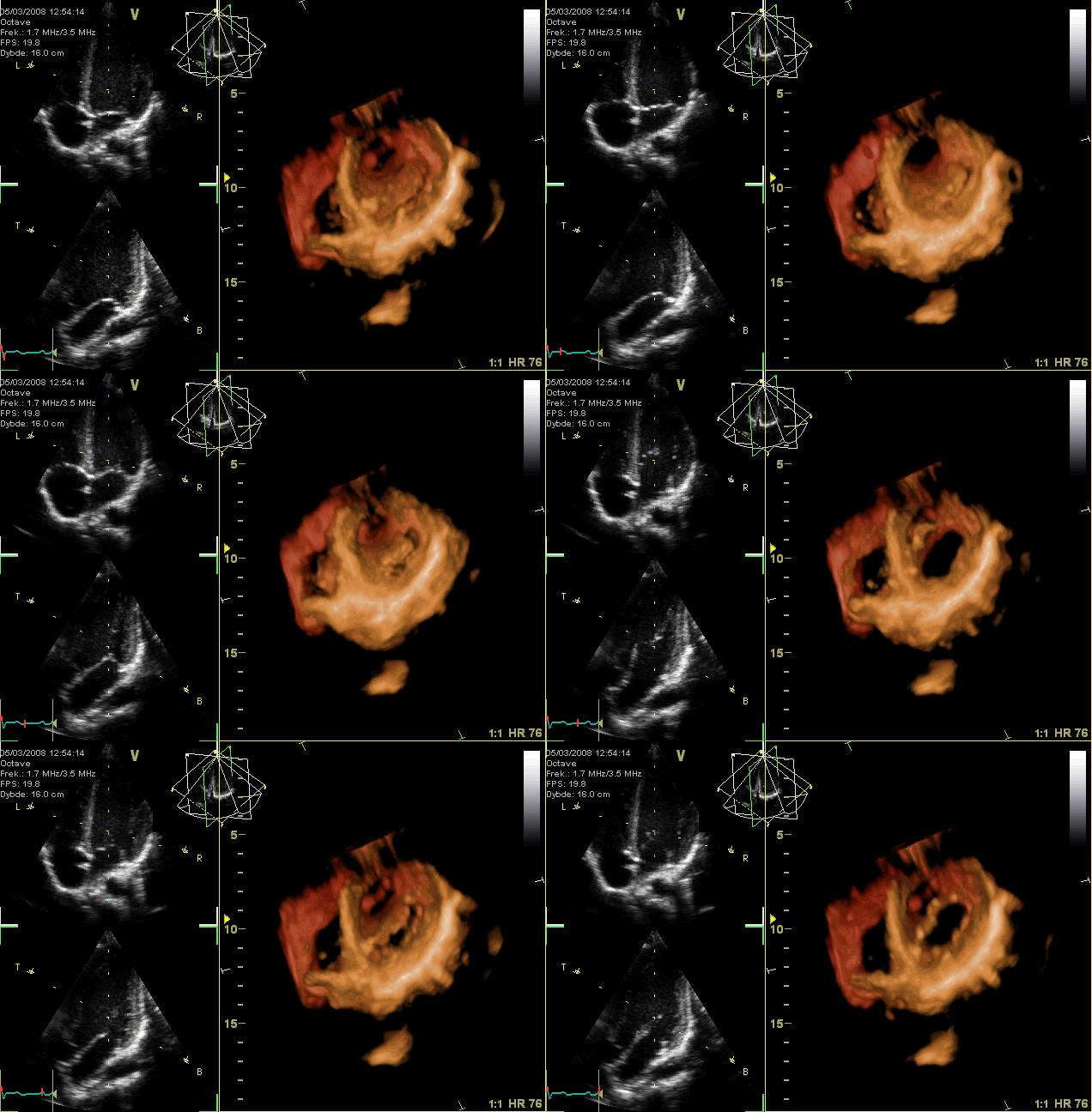
العربية: صُورةٌ مُتحرِّكة لِتخطيط صدى القلب؛ دائرةٌ كaهربائيَّة ثُلاثيَّة الأبعاد للقلب كما يبدو للناظر من الأعلى، حيثُ تمَّت إزالة الجُزء القمّي من
البطينين بينما يظهر الصمَّام التاجي واضحًا للعيان. لا يظهر الصمَّامان الأبهر وثُلاثيّ الشرف بشكلٍ واضح نتيجة ضياع البيانات الخاصَّة بهذا التصوير، بينما تظهر الفتحات. يظهرُ على اليسار منظران ثُنائيَّا الأبعاد مأخوذان بناءً على بيانات الرسم ثُلاثي الأبعاد. يُفسِّرُ
هذا الرسم الصُورة المُتحرِّكة.
English: GIF-animation showing a moving echocardiogram; a 3D-loop of a heart viewed from the apex, with the apical part of the ventricles removed and the mitral valve clearly visible. Due to missing data the leaflet of the tricuspid and aortic valve is not clearly visible, but the openings are. To the left are two standard two-dimensional views taken from the 3D dataset.
Why might echocardiogram imaging be helpful?
An echocardiogram is a non-invasive procedure used to evaluate the functionality and structure of the heart. This kind of imaging might be helpful as it allows the examination of the heart's anatomy and the blood vessels around it (NHS, 2022). The procedure also analyses how blood flows through the veins and evaluates the heart's pumping chambers. In addition, it aids in diagnosing and monitoring some cardiac diseases.
What kinds of issues or anomalies might an echocardiogram imaging be able to detect?
An echocardiogram might detect atherosclerosis, where fatty molecules and other substances in the bloodstream gradually block the arteries. This heart condition may result in issues with the heart's pumping or wall movements (Johns Hopkins Medicine, 2022). The procedure can also detect cardiomyopathy, a cardiac enlargement brought on by thick and frail heart muscle. Additionally, congenital heart defects that appear in one or more cardiac components while a fetus is still developing might be detected using this procedure (Hopkins Medicine, 2022). Moreover, failure of the heart might be noticed. This condition happens when blood cannot be pumped effectively because the heart muscle has grown weak or tight due to cardiac relaxation. Some symptoms of this condition include swelling in the feet, ankles, and other areas of the body and fluid accumulation in blood vessels and lungs.
Furthermore, aneurysm and heart valve malfunction might be detected. An aneurysm is an enlargement and weakening of the aorta or a portion of the heart muscle. If this condition occurs, there is a chance that the aneurysm will burst. On the other hand, heart valve malfunction involves the failure of one or more heart valves, which could result in irregular blood flow within the heart. The constriction of the valves prevents adequate blood flow. As a result, blood might ooze backward through the defective valves, which is dangerous (Johns Hopkins Medicine, 2022). Therefore, using an echocardiogram, heart valves can be examined for infection. At the same time, doctors can determine the best treatment for these diseases.
https://www.intechopen.com/chapters/44904
What might an echocardiogram not be able to detect?
An echocardiogram cannot reveal if one has blocked or clogged arteries. According to Discoverecho (2019), echocardiography is not a medical test particularly good at finding blocked arteries. This setback is mainly due to coronary arteries being typically too tiny to be seen using echocardiography.
References
Discoverecho. (2019, Nov 13). Does an echocardiogram show blockages? (Blocked arteries). https://discoverecho.com/does-echocardiogram-show-blockages/
Jamil, G., Abbas, A., Shehab, A., & Qureshi, A. (2013, Jun 12). Echocardiography findings in common primary and secondary cardiomyopathies. https://www.intechopen.com/chapters/44904
Johns Hopkins Medicine. (2022). Echocardiogram. https://www.hopkinsmedicine.org/health/treatment-tests-and-therapies/echocardiogram NHS. (2022, Mar 28). Echocardiogram. https://www.nhs.uk/conditions/echocardiogram/
A
Sketch explains the animation.
Français : Animation GIF en boucle montrant une échocardiographie en trois dimensions animée. Le cœur est ici vu depuis son apex, avec la partie apicale des ventricules enlevée. La valve mitrale est clairement visible. A cause de la résolution, les feuillets des valves tricuspide et aortique ne sont pas visibles. A gauche se trouvent deux vues bidimensionnelles standard extraites des données tridimensionnelles. L'animation est expliquée par un schéma visible
ici.


